
Answer to Question #11781 Submitted to "Ask the Experts"
Category: Medical and Dental Patient Issues — Diagnostic X Ray and CT
The following question was answered by an expert in the appropriate field:
My daughter recently had elbow radiographs. The settings were 60 kilovolts potential (kVp) and 2.5 milliamps (mA). Her head was only about 0.3 meter (m) away from the tube. I am wondering how much leakage would there be at this distance? I found online that the legal amount of tube leakage would be 1 millisievert (mSv) per hour at 1 m. The tube end had a red line (minus) that was by her head. I want to know what her possible head dose could be from what might have come from the tube. I tried to do the calculation and came up with 16 mSv per second at 0.3 m.
The amount of leakage radiation your daughter was exposed to was miniscule.
Leakage radiation is limited by regulation to 100 milliroentgens per hour (mR h-1) at 1 m.* This is equal to 0.875 milligray per hour (mGy h-1). The maximum leakage is created when the x-ray tube is operated at its highest tube voltage and at a tube current that it can continuously operate. For diagnostic radiographic equipment these are usually 150 kVp and 3–5 mA, respectively. Diagnostic x-ray tubes are shielded such that the actual maximum leakage is between one-half and one-tenth of the limit. At a tube voltage of 60 kVp, the shielding is much more effective. In fact, leakage radiation decreases by more than eight orders of magnitude when comparing leakage at 150 kVp to leakage at 50 kVp. That's 100 million times less. Unfortunately, I do not have ready access to the difference between 150 kVp and 60 kVp, but it should be clear that the dose rate is very, very low.
When we design shielding for an x-ray room, we take leakage into consideration. The average amount of leakage from all types of procedures is used when making this assessment. That average is 0.00014 mGy per patient at 1 m. This includes procedures done at more than 60 kVp, so it is an overestimate of the dose per patient. Now all we need to do is correct for distance. Radiation dose rates decrease with the square of the distance. This means that if you double the distance the dose rate goes down by four, if you triple the distance, the dose rate goes down by nine. In this case, as you go from 1 m to 0.3 m, the dose rate goes up by a factor of approximately 11. The dose per patient is then 0.0015 mGy. Remember, the dose to your daughter's head was less than this.
Finally, we want to convert from mGy to effective dose in mSv. If we make the worst-case assumption that your daughter's entire body and not just her head was exposed to leakage radiation of 0.0015 mGy, then the effective dose would be 0.0015 mSv. With all the worst-case assumptions made in this calculation, her dose from leakage radiation was easily 10 to 100 times less than this.
For perspective, one receives an effective dose from radiation sources in nature of about 0.008 mSv each day one lives on earth. That's five times the overestimate made above. And health effects from radiation exposure have not been seen below 100 mSv.
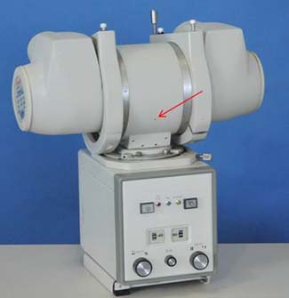
The small line that you described could be the indicator of the position of the focal spot (where x rays are generated). In the pictures below of two different x-ray units, the cylindrical part is the x-ray tube housing, the cube below it is the collimator which is used to adjust the size of the beam, and the red arrow points to the focal spot indicator.

Kent Lambert, CHP, FHPS
* The limit for leakage radiation is given here in units of mR h-1 (called traditional units) because that is the unit used by the regulatory agency. However, the Health Physics Society has adopted the SI (International System) of units, and these units are given in the subsequent sentence.